Uses of Superconductivity in Medical Science
Brief History of Superconductivity
The beginnings of superconductivity can be traced back to the research done by scientist Kammerling Onnes in the year 1911. Onnes, while serving a professorship of physics at the University of Leiden (in the Netherlands), was studying the electrical properties of pure metals at low temperatures (near zero degrees Kelvin) using another recently discovered phenomenon, liquefied helieum. He found that when he immersed the metals (mercury, for historical purposes) the electrical resistivity for the metal suddenly dropped to zero. This was not what was expected, neither by Onnes nor his peers at the time, and was thus the discovery of the phenomenon known as superconductivity.
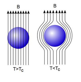
Jump forward to the year 1933, and into the research of Walther Meissner and Robert Ochsenfeld. The two were conducting research in the newly maturing area of metallic properties at low temperatures when they discovered an important characteristic for superconductors (Type I only), the Meissner Effect. The researchers noticed that once their metallic sample had been cooled to a temperature in which it could make its transition into a superconducting state, it would repel any applied magnetic fields that were placed in the sample’s presence. Building upon the work of Meissner and Ochsenfeld, researchers Fritz and Heinz London produced some theoretical groundwork that was useful for explaining the experimental results that had been observed thus far, but little else. Another attempt to explain superconductivity was made in 1950, by scientists L. D. Landau and Vitaly Ginzburg. Although this theory too did little to fully explain the nature of superconductivity, it did predict the splitting of superconducting materials into two categories, Type I and Type II superconductors.
With a growing list of theories that could only explain the experimental results that were found in many laboratories and little else, it was appearing as though superconductivity at the microscopic level would remain a mystery. However, in 1957, researchers John Bardeen, Leon Cooper, and John Schrieffer put forth their work, called the BSC Theory of superconductivity, which provided theoretical groundwork that did more than just explain experiments.
In the last few decades there have been fewer advances to the area of superconductivity. Many hours and many researchers have sought to find so–called high temperature superconductors, but functioning superconductors at room temperature are still in the horizon. With the dearth of understanding of superconductivity mounting, researchers have turned to applications of superconductivity as a means to advance the field.
Applications to Medicine
MRI (Magnetics Resonance Imaging)
Origins
MRI, or Magnetic Resonance Imaging, is probably the most well known application of superconductivity in medicine. It was discovered in 1937 by scientist Isidor Isaac Rabi, who had been studying the effect of magnetic fields on a beam of lithium chloride molecules, a method called molecular beam resonance. Rabi had postulated that with precisely the right perturbation, the magnetic dipole moments of the particles in an atom could be induced to flip their orientation, and when radio waves were passed over the lithium chloride beam, his hypothesis was confirmed. And thus the precursor to the MRI was born, molecular beam magnetic resonance.
The next step towards the modern MRI came from research groups led by Edward Purcell and Felix Bloch at Harvard and Stanford, respectively. Both researchers were studying the nuclear resonance that Rabi had pioneered, but were focusing on hydrogen due to its large magnetic dipole. Each group was able to reproduce the results from Rabi’s experiments using different sources of hydrogen in 1945. The work of Purcell and Bloch is significant to the MRI of today because it showed the practicality of tracking hydrogen using nuclear magnetic resonance. Since hydrogen is so prevalent in the human body, many chemical processes and actions are able to be mapped, and consequently studied.
The final key step in the evolution of the modern day MRI was made back in the late 1940s by two independent researchers, Henry Torrey and Erwin Hahn. Both scientists were able to gather much more complete data on the relaxation time (the time between flips of a particle’s spin) when they switched from a continuous supply of radio waves to a few short pulses of radio waves. These pulses allowed for magnetic resonance imaging to be possible, albeit after a more sophisticated pulse method was developed by Russle Varian, called Fourier transform NMR (Nuclear Magnetic Resonance).
How it Works
As mentioned before, particles such as electrons, protons, or neutrons, all have this thing called a magnetic dipole moment which arises from the quantum mechanical properties of these particles. The magnetic dipole acts like a tiny bar magnet, with a North and South Pole, and consequently produces its own tiny magnetic field. When the atoms are exposed to an external magnetic field, the dipoles of the particles tend to line up with the direction of the field, either in a parallel or anti-parallel fashion, depending on the energy associated with each orientation. However, the “lining up” of the dipole moments in the atoms is a little misleading because the dipole moments do not actually line up directly, but instead precess about the axis of the orientation of the spin (dipole moment). The formal name for this phenomenon is the Larmor Precession and the frequency of the precession is known as the Larmor Frequency. If an outside stimulus can be made to match the Larmor Frequency of the particle, then the particle will absorb enough energy to flip the orientation of its spin and vice versa when the particle releases energy. To accomplish the task of flipping the spins of particles, radio waves employed. By varying the frequency of the radio signals, one can produce flips in the nucleus of different elements.
The magnetic fields that are required for MRI machines need to be strong and uniform (for the most part), so superconductivity was tapped in order to make these a reality. Superconducting magnets create near spatially uniform magnetic fields to within a few ppm (parts per million), as well as large magnetic fields that are stable with time. With the large uniform field set up by the superconducting magnet, another weaker magnetic is superimposed onto already present field, creating a magnetic field gradient. This gradient in the field is necessary for precisely determining the positions of all the different nuclei’s spin. Patients are placed so that the area under consideration is in the magnetic field where it may then be bombarded with radio signals. Once the correct radio frequency has been attained, flips will be induced in the nuclei in the region under investigation. The energy given off from the flips is then read by a computer that runs the data through a program for the Fourier analysis that transforms the different frequencies into an image that can be viewed by a physician. Because the nuclei of different elements have their own intrinsic magnetic resonance frequency, the images produced from an MRI scan give great insight into what is in a given area of the body and what’s going on there, chemically.
With this technique, doctors and researchers are able to get a view of the microscopic workings of the human body (or any material) without being too invasive. MRI machines can be used for functions such as mapping chemical activities in the brain or detecting cancerous tumors. Even the base idea for MRI machines, NMR, requires the precision brought by superconducting magnets. For organic chemists, NMR is an invaluable tool for investigating the organic synthesis of molecules used in pharmaceuticals all the way to the polymers that make up many widely used plastics
